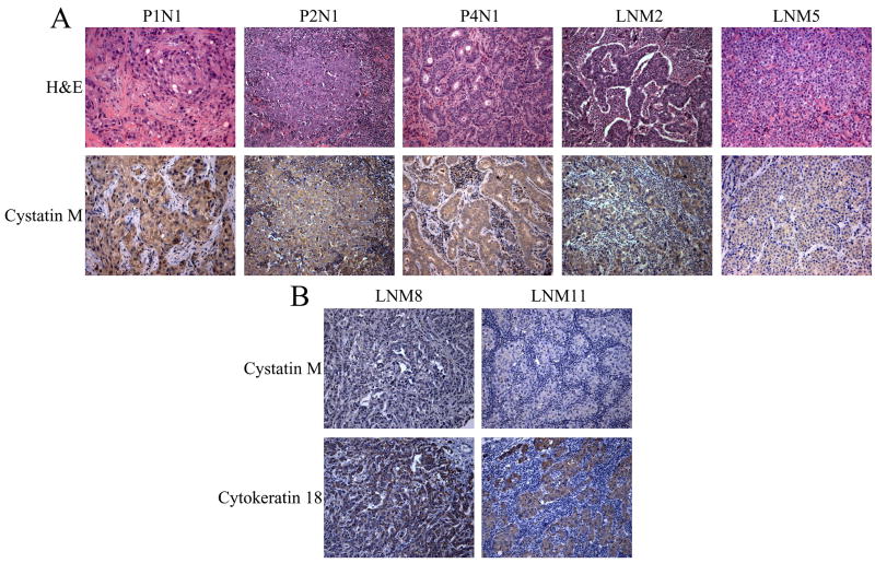
Fig. 2.
Immunohistochemical analysis of cystatin M expression in lymph node metastases. (A) Panels show H&E and cystatin M immunostaining in the same lymph nodes. Lymph node P1N1 shows positive staining for cystatin M. Lymph nodes P2N1, P4N1, LNM2, and LNM5 show reduced cystatin M immunostaining. (B) Panels show cytokeratin 18 (CK18) and cystatin M immunostaining in the same lymph nodes. All metastatic lesions show strong staining for CK18 and exhibit reduced cystatin M immunostaining. (Original objective lens magnification 10x).

