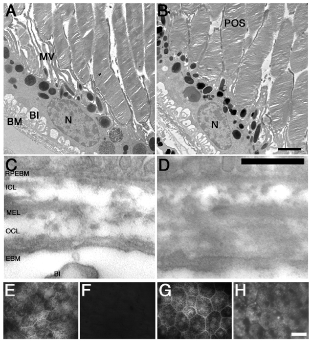
Figure 2.
Age-related changes in F1 F344BN hybrid rat RPE. Observation of young (3–4 month-old, A) and aged (24–25 month-old, B) F1 F344BN hybrid rats reveals several of the RPE age-related changes previously described. These include: Bruch’s membrane thickening (D), accumulation of residual bodies, and microvilli atrophy (B). In addition, bright-field analysis of aged RPE whole-mounts reveals decrease in RPE density (G) while epifluorescence in the green channel (FITC filter: excitation 495 nm/emission 519 nm) reveals increased lipofuscin accumulation (H) when compared to the young RPE cells (E and F). A–D. Transmission electron microscopy.
Abbreviations: BI, basal infoldings; MV, microvilli; POS, photorecptor outer segments; RPEBM, RPE basement membrane; ICL, inner collagenous layer; MEL, middle elastic layer; OCL, outer collagenous layer; EBM, choroidal endothelial cell basement membrane; Bars: (A and B), 1 μm; (C and D), 2 μm; and (C to F), 200 μm.

