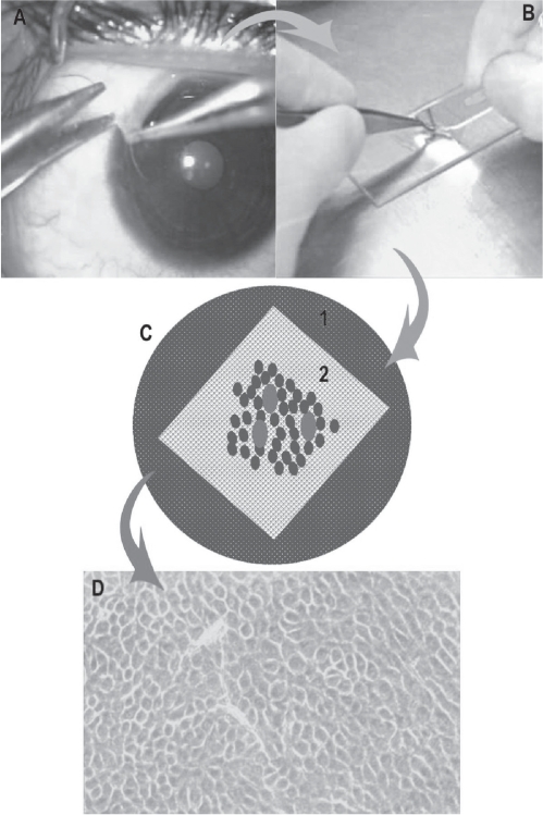
Figure 4.
Schematic diagram showing the steps in cultivation of limbal epithelial stem cells. A: Technique of limbal biopsy (See text for details). B: Processing of tissue in the laboratory and making the explant culture. C: Petridish, glass slide (white) with de-epithelialized human amniotic membrane with explants (red dots) with growing cells around it (blue dots). D: Monolayer of cells (10–14 days old) under phase contrast microscope.

