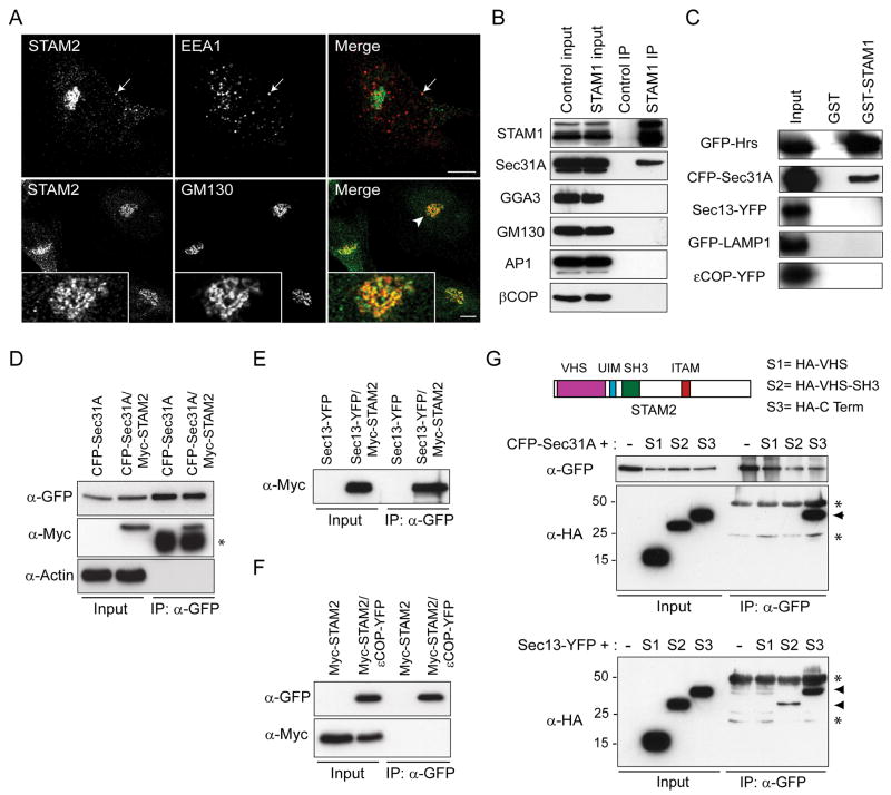
Figure 1. STAMs interact with proteins of the early secretory pathway.
A) HeLa cells were co-immunostained for endogenous STAM2 (green) and either EEA1 or GM130 (red), then visualized using confocal microscopy. Arrows in the upper panels identify an area of colocalization, and an arrowhead in a lower panel identifies an area enlarged in the insets. Bar, 10 μm. B) HeLa cell lysates were immunoprecipitated (IP) with anti-STAM1 antibodies or control IgG and immunoblotted with antibodies against endogenous STAM1, Sec31A, GGA3, GM130, AP1, or βCOP. Inputs represent 5% of the starting material. C) Lysates from cells expressing the indicated constructs were incubated with either GST or GST-STAM1 immobilized on glutathione-Sepharose beads and immunoblotted with anti-GFP antibodies. Inputs represent 5% of the starting material. D and E) HeLa cells were transfected with CFP-Sec31A (D) or Sec13-YFP (E) or else co-transfected with either construct and Myc-STAM2. Lysates were immunoprecipitated with anti-GFP antibodies and immunoblotted with antibodies against GFP or Myc-epitope. Both Sec31 and Sec13 co-precipitate with STAM2. Actin was probed to monitor specificity. An asterisk (*) in (D) denotes the IgG heavy chain. F) HeLa cells were transfected with Myc-STAM2 or else co-transfected with Myc-STAM2 and εCOP-YFP, then immunoprecipitated with anti-GFP antibodies. G) HeLa cells were singly transfected with CFP-Sec31A or co-transfected with CFP-Sec31A and HA-tagged STAM2 domains (schematic diagram on right). Cell lysates were immunoprecipitated with anti-GFP antibodies and immunoblotted with anti-GFP or anti-HA antibodies (left panels). Only the C-terminal region of STAM2 co-precipitates with Sec31 (arrowhead); asterisks (*) denote IgG heavy and light chains. HeLa cells were also singly transfected with Sec13-YFP or co-transfected with Sec13-YFP and HA-tagged STAM2 domain constructs, then immunoprecipitated with anti-GFP antibodies (middle panels). Both VHS-SH3 and C-terminal segments of STAM2 co-precipitate with Sec13 (arrowheads); asterisks (*) identify IgG heavy and light chains.

