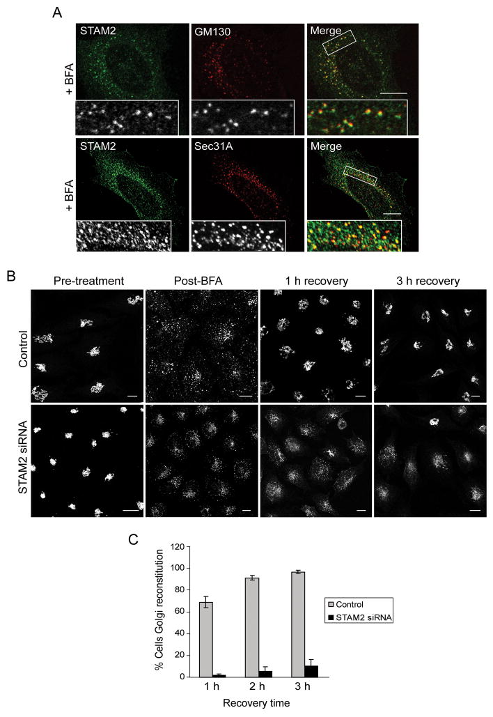
Figure 7. Localization of STAM2 to ERES in BFA-treated cells and delayed recovery of Golgi morphology in STAM2 siRNA cells following BFA.
A) HeLa cells were treated with BFA and co-stained for STAM2 (green) and GM130 or Sec31A (red). STAM2 puncta are adjacent to GM130 puncta, and STAM2 puncta colocalize with Sec31A puncta. B) HeLa cells transfected with control or STAM2 siRNAs were treated with BFA and assessed after wash-out. Most control cells (upper panels) display normal Golgi morphology as assessed by GM130 staining after 1 h of recovery, whereas most STAM2 siRNA-treated cells (lower panels) have not recovered even after 3 h. Bar, 10 μm. C) Graphical presentation of the percentage of cells with Golgi reconstitution (lacking tubular Golgi) after transfection with STAM2 siRNA and control siRNA (n=3; 100 cells per experiment; ±SD).

