
FIG. 5.
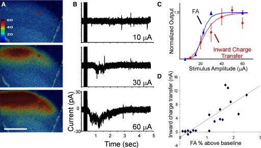
A: FA in somatosensory cortex in response to VPM stimulation at 10, 30, and 60 μA. B: current changes in a cell at the location of the red spot held in voltage clamp at −60 mV. The traces represent the mean of 10 runs. Vertical lines on the left of the traces represent the stimulus artifact. C: normalized mean input–output functions from 4 cells (inward charge transfer, red) and their corresponding ROIs (increase in fluorescence in 3 × 3-pixel areas around cell, blue). Fitted function is a Naka–Rushton function (see methods) and error bars represent normalized SE. D: scatterplot of FA amplitude (expressed as percentage increase from baseline) vs. total inward charge transfer for all stimulus amplitudes across 4 neurons. R2 = 0.67, t = 0.001, t = 6.02, linear fit: y = 5.0x − 1.6. Stimulus parameters: monopolar electrical stimulation using 2-ms-duration pulses, 40 pulses/s for 250-ms train, 14-s intertrain intervals for a total of 2 pulse trains, and 28 s of imaging time.