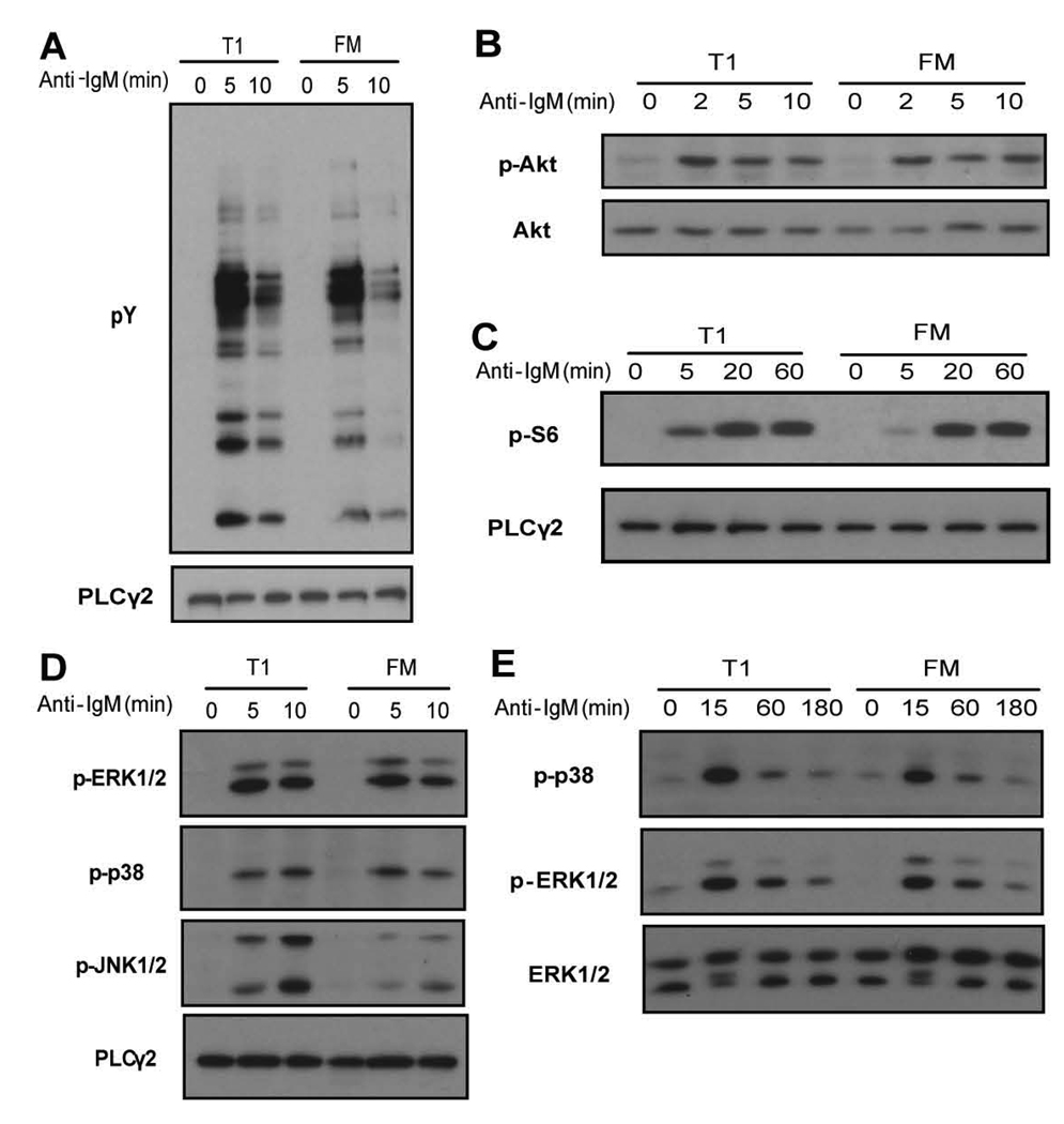
Figure 3.
Activation status of proximal signaling molecules in sorted T1 and FM B cells upon anti-IgM stimulation. Whole cell lysates from stimulated and unstimulated cells were probed with antibodies that recognize: A, total pY B, phosphorylation of AKT at S473 C, phosphorylation of S6 D, phosphorylation of ERK1/2, p38 or JNK1/2 (short time points) or E, ERK1/2 and p38 (longer time points). Blots were stripped and reprobed with anti-PLCγ2, anti-AKT or anti-ERK1/2 to evaluate protein loading. Data shown are representative of at least 3 independent experiments.

