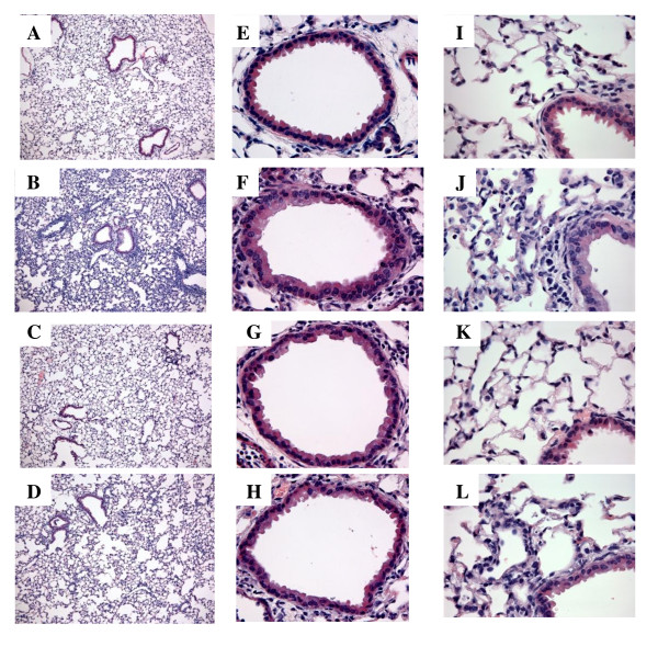
Figure 2.
TLR3 KO mice are partially protected from poly(I:C)-induced inflammation in lung interstitium. Representative H&E-stained lung sections from WT- PBS treated (A,E, I)WT poly(I:C)-treated (B, F, J), TLR3 KO PBS treated mice (C ,G, K) and TLR3 KO poly(I:C)-treated (D, H, L). Figures A-L are representative images from each group. Figure A-D are at 10×, Figures E-H are at 40 × and Figures I-L are at 60 ×.

