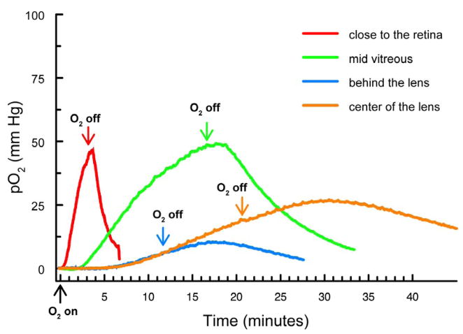
Figure 2.
Increase in pO2 levels in various regions of vitreous humor and in the lens center of normal guinea pigs after breathing 100% O2. Arrows indicate the start of 100% O2 exposure and a return to breathing room air. red: close to the retina; green: mid-vitreous; blue: directly behind the lens; orange: center of the lens.

