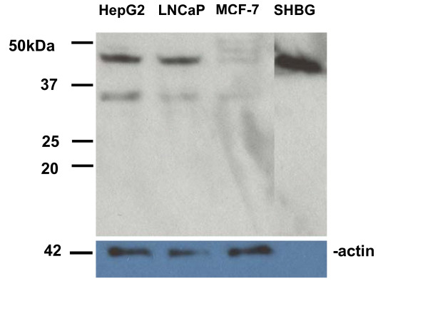
Figure 8.
Western blot analysis of SHBG protein expression in HepG2, LNCaP, and MCF-7 cells. Protein extracts (20 μg each) prepared from HepG2, LNCaP, and MCF-7 cells, and 10 picograms of purified SHBG protein isolated from human plasma using a steroid affinity column, were electrophoresed through 10% LongLife polyacrylamide gels (Gradipore-VWR), transferred to PVDF membranes, and hybridized to either a rabbit anti-human SHBG polyclonal antibody (top, WAK-S1012-53, WAK-Chemie, Germany) or a rabbit anti-actin affinity purified polyclonal antibody (bottom, A2066, Sigma). Molecular weight (in kilodaltons) marker positions are given on the left. The top band in HepG2 and LNCaP cells migrates with a molecular weight of approximately 44–46 kD, while the bottom band migrates with a molecular weight of approximately 33–35 kD. The two larger bands in MCF-7 migrate with molecular weights of 51–53 kD and 48–50 kD, respectively. Note- the size marker appears to have run a bit slowly on the top gel, as the purified SHBG is slightly smaller than prior reports of 50–52 kD.

