
Figure 1. Elastin distribution in en face sections of dermis and desmosine analysis.
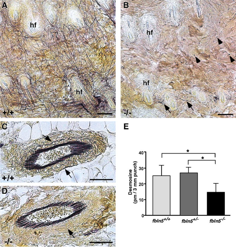
In 2 month-old wild-type skin (A), an extensive elastic fiber network (black lines) can be seen between the hair follicles (hf). In fibulin-5 null skin (B), only a few intact elastic fibers (arrows) can be seen near the hair follicles (hf). Distant to the hair follicles, very small foci of elastin-positive staining are evident (arrowheads). In contrast to the dermis, small blood vessels near the hypodermis appear similar between wild-type (C) and fibulin-5 null (D) skin. Closer examination, however, does show a difference in the elastic fiber distribution in the surrounding media and adventitia (C, D - arrows). Desmosine analysis of 14 day-old mouse skin shows a significant decrease in amount of desmosine, a measure of mature cross-linked elastin, in Fbln5−/− skin compared to wild-type or Fbln5+/− skin (means ± S.D.; * = p < 0.05; n = 5 for each genotype). Scale bars = 50 μm.