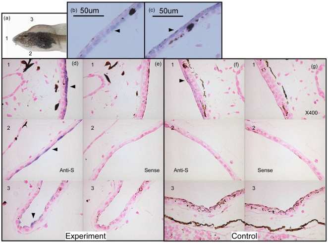
Figure 5. In situ hybridization (ISH) analysis of control and bulgy morph tadpoles.
(a) Tadpoles were longitudinally sectioned (5 µm) at the frontal side (1), the ventral side (2), and the dorsal side (3). (b) ISH analysis of bulgy tadpoles with an anti-sense probe for type-1 collagen expression. Arrowhead indicates type-1 collagen signal. (c) ISH of bulgy tadpoles with an anti-sense probe for pirica expression. Arrowhead indicates the signal for pirica expression. (d) Pirica gene expression with an anti-sense probe at the frontal side (1), ventral side (2), and dorsal side (3) of a bulgy morph tadpole. (e) ISH of pirica gene expression using a sense probe. (f) ISH of pirica gene expression in a control tadpole using an anti-sense probe. (g) ISH of pirica gene expression in a control tadpole using a sense probe.

