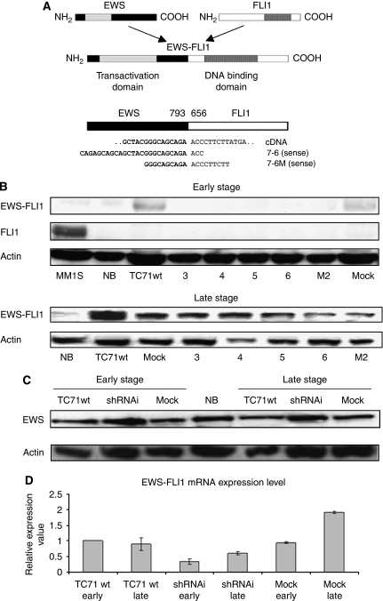
Figure 1.
Targeting of EWS–FLI1 through shRNAi for stable protein and mRNA knockdown. (A) Diagram of wild-type EWS, wild-type FLI1, and EWS–FLI1 fusion type 1. The illustration of EWS–FLI1 cDNA and the location of the two shRNAi designs used, 7-6 and 7-6M, both against fusion break-point. (B) Analysis by western blotting showed that the EWS–FLI1 expression was reduced in several TC71 shRNAi clones. The EWS–FLI1 reduction is maintained partially in some shRNAi clones through the course of cellular passages (early and late stages). EWS–FLI1 68 kDa, FLI1 51 kDa, and actin 42 kDa. 3, 4, 5, 6: TC71 shRNAi clones transfected with shRNAi design type 7-6; M2: TC71 shRNAi clone transfected with shRNAi design type 7-6M. MM1S: multiple myeloma cell line. Positive control for FLI1. NB: neuroblastoma cell line SK-N-JD. Negative control for EWS–FLI1. Actin is shown as a loading control. (C) Analysis by western blotting showed that the EWS–FLI1 shRNAi designs are specific and did not alter the EWS expression in the early and late stages. The shRNAi clone corresponds to the TC71 shRNAi clone 6. Actin is shown as a loading control. (D) The shRNAi reduction in the EWS–FLI1 mRNA level assessed through qRT–PCR (SYBR green probes) in the early (T0) and late passage (T8). The shRNAi clone corresponds to the TC71 shRNAi clone 6. GAPDH was used as an internal control. Columns, mean of triplicates of three different replicates; bars, s.d.

