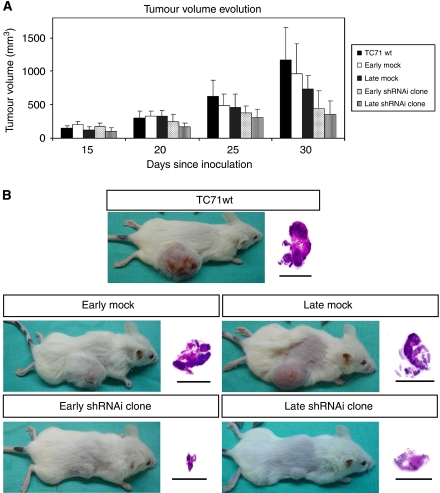
Figure 3.
Tumour growth evolution. The EWS–FLI1 shRNAi reduced tumoural growth in vivo. (A) NOD-Scid mice were s.c. injected with 5 × 10⁶ TC71 cells. The treated mice (early and late shRNAi clone) showed smaller tumours than the control groups (TC71wt, late and early mock) since the third week after cells injection. At the end of the study these differences were statistically significant (P<0.05). (B) Visual and histopathological evaluation of mice tumours. All of the tumours showed the same histopathological pattern: a large area of necrotic tissue, an area of proliferating cells, and a layer between them with cells in apoptosis.

