Figure 4.
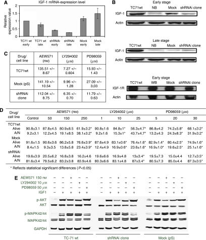
EWS–FLI1 shRNAi impaired the IGF-1/IGF-1R survival pathway and its downstream targets. (A) Histogram representation of IGF-1 transcript level knockdown in the shRNAi clone as assessed by qRT–PCR normalised to GAPDH. The shRNAi clone corresponds to the TC71 shRNAi clone 6. SYBR probes were used. All experiments were performed in triplicate in the early and late stages. Columns, mean of triplicates of three different experiments; bars, s.d. (B) The IGF-1 protein level is reduced in the shRNAi clone as assessed through western blot whereas the IGF-1R protein level is not changed. The shRNAi clone corresponds to the TC71 shRNAi clone 6. Actin is shown as loading control. (C) IC50 of proliferation measured by MTT assay after the NVP-AEW541, LY294002, or PD98059 treatment (72 h of incubation). The shRNAi clone showed more sensitivity to the action of inhibitors of the IGF-1/IGF-1R pathway being statistically significant for AEW571 and PD98059 (P<0.05). The shRNAi clone corresponds to the TC71 shRNAi clone 6. (D) Apoptotic index after the NVP-AEW541, LY294002, or PD98059 treatment (72 h of incubation). Apoptosis could be further induced in the shRNAi clone under all conditions. The means±standard deviations (error bars) of four independent experiments are shown. The shRNAi clone corresponds to the TC71 shRNAi clone 6. P<0.05 values are considered as significant. (E) Effects of NVP-AEW541 combined with inhibitors LY294002 and PD98059 on the activation of the IGF-1/IGF-1R signalling pathway. The AKT and MAPK42/44 phosphorilation were diminished in the shRNAi clone under IGF-1 stimulation whereas TC71wt and mock cells maintained the active phosphorylation status in both kinases. All conditions were treated with AEW571 during 15 min and with the inhibitors for 2 h, before IGF-1 stimulation (50 ng ml−1) during 15 min (serum-free conditions). The shRNAi clone corresponds to the TC71 shRNAi clone 6.

