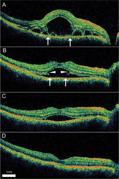
Fig. 3.
Serial optical coherence tomography (OCT) scans from a 32-year-old man diagnosed with Vogt-Koyanagi-Harada disease. (A) Intraretinal fluid is noted on a 9 mm horizontal OCT scan of the right eye after three days of treatment with oral prednisolone. The margins of the cystoid space are indicated by arrows. (B) Methylprednisolone (125 mg) was infused intravenously for three days. On day six, as the outer boundary of the intraretinal space degraded, the cystoid and the subretinal spaces became interconnected (arrows). Defects or notches are seen at the margins of the previous cystoid space (arrowheads). An oral steroid was prescribed again. (C and D) The subretinal fluid gradually resolved over one month.

