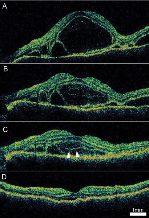
Fig. 4.
Serial 9 mm vertical optical coherence tomography (OCT) scans from the right eye of a 43-year-old woman diagnosed with Vogt-Koyanagi-Harada disease. (A) An OCT scan before steroid treatment demonstrates a large cystoid space involving the fovea (arrows). (B) On the third day, multiple layered structures are noted in the cystoid space. (C) On day seven, the cystoid space decreased in size and the layered structure became more distinct. (D) After two weeks of steroid treatment, a small volume of subretinal fluid was noted under the fovea without a cystoid space.

