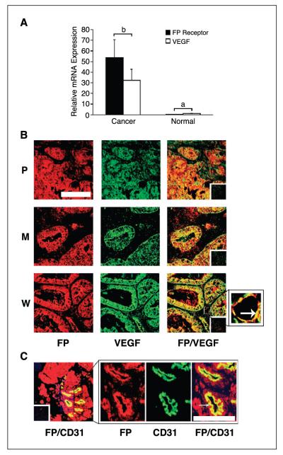
Figure 1.
A, relative mRNA expression of FP receptor and VEGF in endometrial adenocarcinoma (n = 25) and normal endometrium (n = 10) as determined by real-time quantitative RT-PCR analysis. B, localization of the site of expression of FP receptor (red) and VEGF (green) and colocalization of FP receptor with VEGF (merged; yellow) in poorly (P), moderately (M), and well-differentiated (W) endometrial adenocarcinomas, respectively. Arrow, blood vessel under high magnification. Bar, 100 μm. C, localization of the site of expression of FP receptor (red) and CD31 (green) and colocalization of FP receptor with CD31 (merged; yellow) in the endothelial cells of the blood vessels. Representative sample of a moderately differentiated adenocarcinoma. Insets, negative controls. Bar, 10 μm. P < 0.05, b is significantly different from a.

