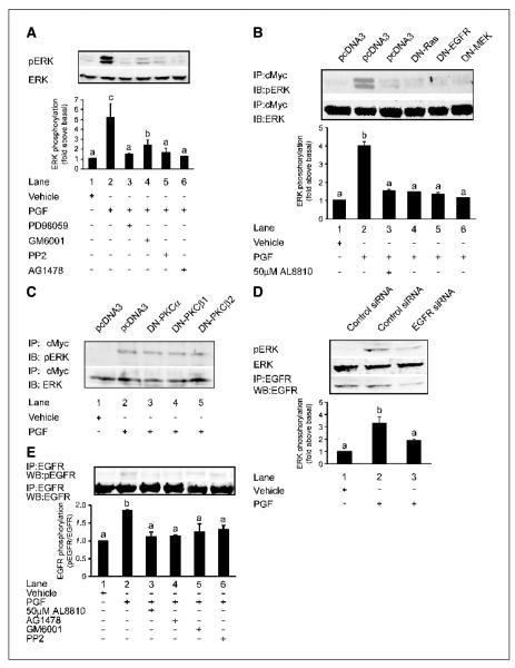
Figure 4.
Representative Western blots showing activation of ERK1/2 and EGFR signaling in FPS Ishikawa cells. A, FPS cells were pretreated for 1 hour with inhibitors or vehicle followed by stimulation with vehicle (control; lane 1), 100 nmol/L PGF2α (lane 2), 100 nmol/L PGF2α and PD98059 (lane 3), 100 nmol/L PGF2α and GM6001 (lane 4), 100 nmol/L PGF2α and PP2 (lane 5), and 100 nmol/L PGF2α and AG1478 (lane 6) for 10 minutes. B, Ishikawa FPS cells were transfected with c-Myc-tagged ERK cDNA together with pcDNA3 (control empty vector) cDNA or cDNA encoding DN-Ras, DN-EGFR, or DN-MEK. Cells were pretreated for 1 hour with 50 μmol/L AL8810 or vehicle followed by stimulation with vehicle (control), 100 nmol/L PGF2α, or 100 nmol/L PGF2α and 50 μmol/L AL8810 for 10 minutes. C, Ishikawa FPS cells were transfected with c-Myc-tagged ERK cDNA together with pcDNA3 cDNA or cDNA encoding DN-PKCα, DN-PKCβ1, and DN-PKCβ2. Cells were stimulated with vehicle (control) or 100 nmol/L PGF2α for 10 minutes. D, Ishikawa FPS cells were transfected with control random siRNA or siRNA targeted against the EGFR. Cells were stimulated with vehicle or 100 nmol/L PGF2α for 10 minutes and subjected to immunoblot analysis for phosphorylated ERK1/2, total ERK, or immunoprecipitated (IP) and immunoblotted (WB) with antisera against EGFR (bottom). E, representative Western blot showing PGF2α transactivation of EGFR signaling in FPS cells. FPS cells were pretreated for 1 hour with inhibitors, antagonist, or vehicle followed by stimulation with vehicle (control; lane 1), 100 nmol/L PGF2α (lane 2), 100 nmol/L PGF2α and AL8810 (lane 3), 100 nmol/L PGF2α and AG1478 (lane 4), 100 nmol/L PGF2α and GM6001 (lane 5), and 100 nmol/L PGF2α and PP2 (lane 6) for 10 minutes. After lysis, EGFR was immunoprecipitated with anti-EGFR antibody and tyrosine-phosphorylated EGFR was detected by immunoblotting with anti-phospho-EGFR antibody (top). The total amount of EGFR in immunoprecipitates was determined by reprobing the same blot with anti-EGFR antibody (bottom). Immunoblots were quantified as described in Materials and Methods. Columns, mean of four independent experiments; bars, SE. P < 0.05, b is significantly different from a; P < 0.01, c is significantly different from a and b. -, ab- sence of agent; +, presence of agent.

