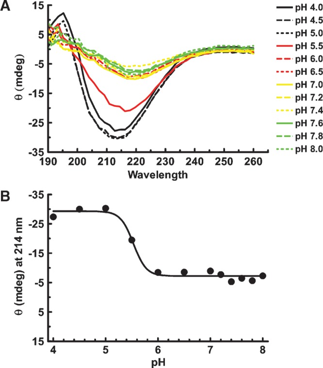
Fig. 3.

pH titration of soluble apoB[37–41] fragment. A: ApoB[37–41] protein was diluted to 14 μM (0.3 mg/ml) in 20 mM phosphate buffer containing 50 mM NaCl at the indicated pH. After incubation overnight at 4°C, the far UV spectrum was collected on an Olis DSM 17 CD spectrophotometer. Compton calculations of secondary structure contributions were performed at pH 3.0 using the instrument software, revealing 15.6% α-helix, 28.1% β-sheet, 24.6% β-turn, and 34.4% other structure. B: Ellipticity (θ) at 214 nm is plotted as a function of pH. Evidence of apoB structure greatly diminished above pH 5.5 due to precipitation of apoB[37–41] at higher pH.
