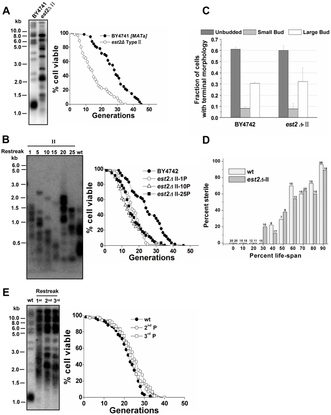
Figure 2. Premature aging of telomerase-null type II survivors.
(A) Telomere analysis (left) and life span assay (right) of telomerase-null type II survivors. The number of daughter cells (generations) produced per mother cell are plotted as a function of mother cell viability. Average life span of strains shown (sample size): BY4741, 27.8±9.7 (n = 60); est2Δ-type II (MATa), 15.3±8.5 (n = 57). (B) Changes of telomeres (left) and life span (right) in type II survivors during the outgrowth. Type II survivors were serially restreaked 25 times on YPD plates. (Left) Genomic DNA from the 1st, 5th, 10th, 15th, 20th, and 25th restreaks was digested with 4 bp cutter (MspI, HaeIII, HinfI, AluI) for the Southern blot analysis. (Right) Life span of the 1st, 10th, and 25th restreaks was determined. Average life span of strains shown: BY4742, 24.8±9.6 (n = 59); est2Δ-type II -1st P, 16.5±6.2 (n = 57); est2Δ-type II -10th P, 15.0±7.9 (n = 57); est2Δ-type II -25th P, 16.1±7.6 (n = 60). (C) Terminal morphology of senescent cells. Cells at the end of life span experiments were classified according to the budding pattern as described [37]. Average and S.D. values from three independent experiments are shown (n>50 for each strain in each experiment). (D) α factor responsiveness in old cells. Cells of various ages were scored for their ability to undergo cell cycle arrest and schmooing in response to the yeast mating pheromone, α factor. The number of cells in each data set for each age group is shown above the bar and data is presented as the percentage of cells that did not schmoo in the presence of α factor. (E) Life span (right) and telomere length (left) analysis. Four telomerase components: Est1p, Est2p, Est3p, and TLC1 RNA were co-overexpressed in BCY123 under the control of GAL1 promoter (Galactose induction). Then cells were serially restreaked on YPD to shut off the over-expression of telomerase. (Left) Telomere blot analysis of the 1st, 2nd, and 3rd passaged cells. Genomic DNA was digested with XhoI. (Right) Life span analysis of the 2nd and 3rd passaged cells. Average life span of strains shown: wild-type, 21.8±6.7 (n = 60); 2nd P, 23.9±7.5 (n = 60); 3rd P, 23.8±6.6 (n = 60).

