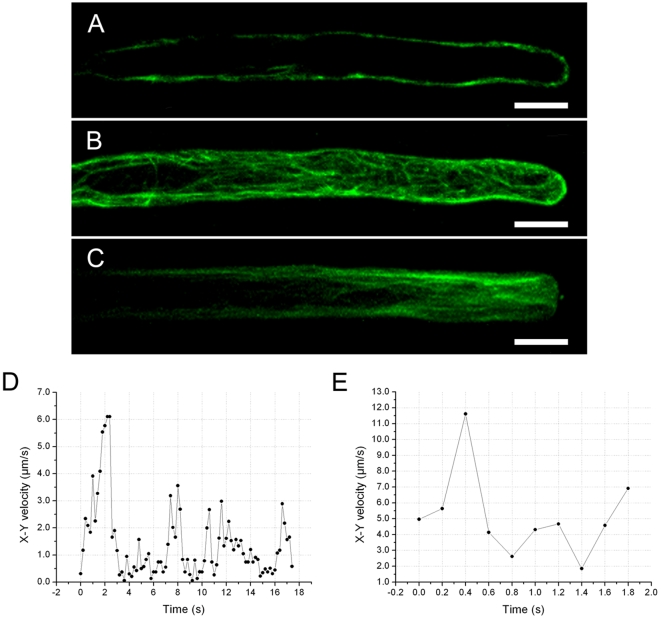
Figure 5. Effects of microtubule-active drugs on actin filaments and mitochondrial movements.
A. A single optical section of interior cytoplasm of the root hair of B. Note that no actin filaments are visible in the interior cytoplasm. Scale bar = 10 µm. B. A growing root hair treated with 10 µM oryzalin displaying dispersed and curved actin bundles around the cortical cytoplasm and in the base of the hair. Scale bar = 10 µm. C. A growing root hair treated with 5 µM taxol showing an increase in fluorescence in the subapical and apical regions and an aggregation of thick actin bundles in the very tip of the hair, concomitant with a decrease in the basal shank. Scale bar = 10 µm. D. Plot of the x-y velocity of a mitochondrion moving in the region 0–80 µm from the tip of a growing root hair treated with 10 µM oryzalin. E. Plot of the x-y velocity of a mitochondrion moving in the region 0–80 µm from the tip of a growing root hair treated with 5 µM taxol.

