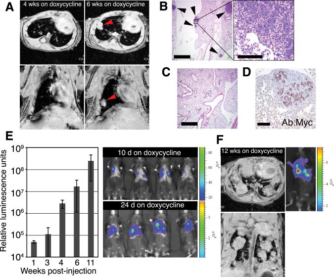
Fig. 1.
Untransformed mouse mammary cells form lung metastases after IV injection and induction of oncogenes. (A to D) Lung metastases develop from intravenously injected phenotypically normal mammary cells upon activation of MYC and KrasD12 transgenes. (A) Metastases were monitored by MRI in Rag1−/− mice after IV delivery of 1 × 10⁶ dissociated primary mammary cells from doxycycline-naïve TOM;TOR;MTB mice. Recipient mice were fed doxycycline for 6 weeks starting 1 day before injection. Representative axial (top) and coronal (bottom) images obtained from the same animal 4 and 6 weeks after injection show development of a solid nodule (arrowheads) in the lung. (B) Foci of hematoxylin-eosin (H/E)–stained mammary adenocarcinoma (arrowheads) in paraffin-embedded lung sections of the same Rag1−/− mouse as in (A). Scale bars indicate 1 mm (left) and 0.1 mm (right). (C) No tumors were observed in lung sections of Rag1−/− mice that did not receive doxycycline after IV delivery of 1 × 10⁶ primary mammary cells from doxycycline-naïve TOM;TOR;MTB mice. Scale bar, 1 mm. (D) Tumor cells from the same animal as in (A), but not the surrounding lung tissue, stained with anti-MYC antisera. Scale bar, 0.1 mm. (E and F) Lung metastases develop from intravenously injected phenotypically normal mammary cells upon activation of a PyMT transgene. (E) Donor cells expressing their transgene were detected by bioluminescence imaging after 5 × 10⁵ primary mammary cells from doxycycline-naïve TOMT:IRES:Luc;MTB mice were injected intravenously into Rag1−/− mice that were placed on doxycycline 1 day before injection. Representative images at day 10 and day 24 after injection show the presence of signal-emitting cells in the thorax (right); temporal increases in bioluminescence (14) were quantified in relative luminescence units (left; n = 5 mice; error bars represent SD). (F) Axial (top) and coronal (bottom) MRI images of a mouse from (E) maintained on doxycycline for 12 weeks show solid nodules in the lung. A corresponding bioluminescence image is shown on the right.

