Fig. 2.
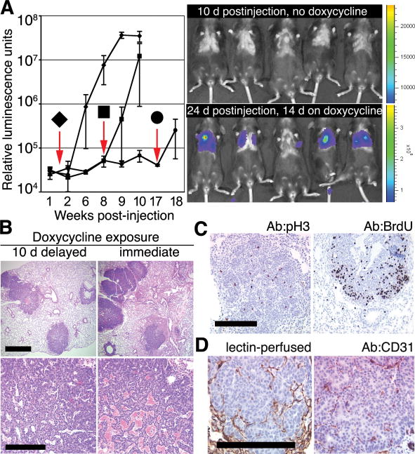
Delay in oncogene activation does not preclude the development of ectopic mammary tumors. (A) Bioluminescence in Rag1−/− mice after IV delivery of 1 × 10⁵ mammary cells from doxycycline-naïve TOMT:IRES:Luc;MTB mice is undetectable before doxycycline exposure but can be induced at various times after placing mice on doxycycline 1.5 (◆, n = 7 mice), 8 (■, n = 3 mice), or 17 weeks (●, n = 2 mice) after IV injection. Downward arrows indicate times of addition of doxycycline to the diet. Error bars represent SD. Representative bioluminescence images (right) obtained 10 days after injection in the absence of doxycycline (top right) and after 2 additional weeks on doxycycline (bottom right). (B) Histologically similar metastatic tumors in lungs of Rag1−/− mice after IV delivery of 1 × 10⁵ mammary cells from doxycycline-naïve TOMT:IRES:Luc;MTB mice exposed to doxycycline for 8 weeks starting 10 days after (left) or 1 day before (right) IV injection. Scale bars, 1 mm (top) and 0.1 mm (bottom). (C) Mitotic activity in tumor foci in lung sections from Rag1−/− recipient of TOM;TOR;MTB cells (left; stained with anti-pH3) or from Rag1−/− recipient of TOMT:IRES:Luc;MTB cells (right; stained with anti-BrdU serum after BrdU labeling) (14). Scale bar, 0.1 mm. (D) Angiogenic proficiency demonstrated by perfusion of the ectopic tumor with biotinylated lectin (left) (14) and by staining endothelial cells within tumor foci with anti-CD31 serum in a Rag−/− recipient of the TOMT:IRES:Luc; MTB cells (right). Scale bar, 0.1 mm.

