Fig. 3.
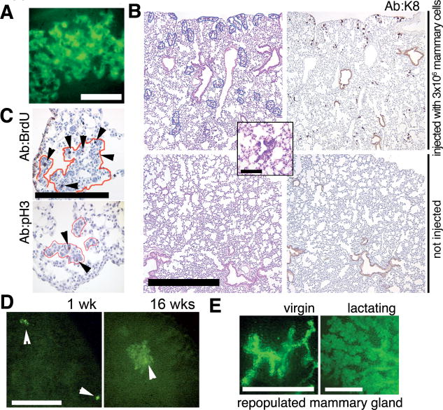
Mammary cells without an oncogenic transgene can persist in the lung. (A) Focus of green cells observed under excitation light (14) in a whole fresh lung of a Rag1−/− recipient 3 weeks after IV injection of 5 × 10⁵ dissociated mammary cells from a β-actin-GFP mouse. Scale bar, 0.1 mm. (B) Representative size and distribution of the H/E–stained ectopic foci in lung sections from Rag1−/− mice injected intravenously with 3 × 10⁶ mammary cells from a β-actin-GFP donor at 3 weeks after injection (top left). Inset shows a representative H/E–stained focus at high magnification. A consecutive section (top right) was stained with rat antiserum against K8. No foci were detected by H/E (bottom left) or K8 staining (bottom right) of lung sections from uninjected Rag1−/− mice. Scale bar, 1 mm. Inset scale bar, 0.2 mm. (C) Mitotic activity in ectopic epithelial outgrowths (outlined in red) demonstrated by BrdU-labeling detected with rat anti-BrdU serum (arrowheads, top) or by staining with anti-pH3 (arrowheads, left). Scale bar, 0.2 mm. (D) Larger foci of green fluorescent cells were observed by whole-lung imaging under excitation light (14) at 16 weeks after injection (right) as compared with 1 week after injection (left). Rag1−/− recipients were injected with 3 × 10⁵ dissociated mammary cells from a β-actin-GFP mouse. Scale bar, 1 mm. (E) Mammary gland repopulation in secondary Rag1−/− recipients produces a green fluorescent mammary tree detectable under excitation light in the whole-mount preparations 4 or 7 weeks after transplantation (14). Glands were harvested from virgin recipients (left), or host animals were mated and the transplanted glands harvested 1 day postpartum (right). Scale bar, 1 mm.

