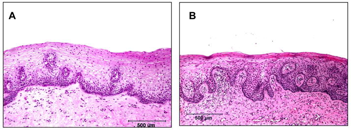
Figure 1.
Comparative histology of the vaginal mucosa of a human (A) and rhesus macaque (B). Note the similarities in the thickness and the arrangement of the the squamous epithelium. Vaginal biopsies were obtained from a normal woman in the luteal phase of the menstrual cycle prior to initiating a study to examine the effects of progestins on the vaginal epithelium (45) and from a normal rhesus macaque in the luteal phase.

