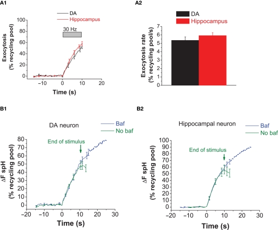
Figure 4.
The rate of exocytosis with a 30 Hz stimulus is similar in DA and hippocampal neurons. (A1) Average exocytosis at 30 Hz in DA (black) and hippocampal (red) neurons. The increase in fluorescence with 30 Hz activity was measured in the presence of bafilomycin. There is close overlap between the exocytosis traces in DA and hippocampal neurons during activity at 30 Hz. Data averaged from six DA and eight hippocampal neurons. Each experiment consists of 10–50 presynaptic terminals from one neuron. Error bars are SEM. (A2) The rate of exocytosis at 30 Hz [slope from linear fitting of traces from (A1)] is similar between DA (black bar) and hippocampal (red bar) neurons. (B1) The average spH response in DA neurons to a 30 Hz stimulus in the absence (green) or presence (blue) of bafilomycin. Green arrows mark the end of the stimulus in the absence of bafilomycin. In the presence of bafilomycin, the stimulus was applied for 120–150 s. The difference between the ‘baf’ and ‘no baf’ traces represents vesicles that have been both internalized and acidified. Data averaged from five DA neurons. Error bars are SEM. (B2) The average spH response in hippocampal neurons to a 30 Hz stimulus in the absence (green) or presence (blue) of Bafilomycin. Data averaged from eight hippocampal neurons. Error bars are SEM.

