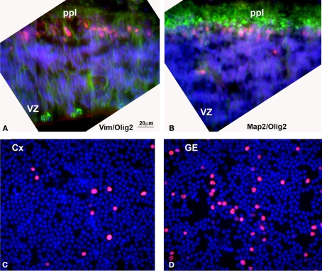
Figure 6.
(A, B) Olig2 expression in human embryonic (5 gw) forebrain. (A) Olig2 cells (red) in the primordial plexiform layer (ppl), but not in the ventricular zone (VZ), where vimentin+ radial glia cells (green) are dividing. (B) Olig2 is expressed in nuclei of MAP2+ young neurons (green) at this stage. (C, D) Densities of Olig2+ cells (red) in the acute (4 h in vitro) dissociated cell cultures from human cortical (Cx) and ganglionic eminence (GE) ventricular/subventricular zones at midgestation (20 gw). Nuclei are counterstained by bisbenzamide (blue). Detailed quantification was published by Jakovcevski and Zecevic (2005b).

