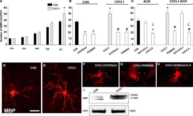
Figure 9.
Effect of CXCL1 on the differentiation of oligodendrocyte progenitors in slice cultures from VZ/SVZ region at midgestation. (A) Time-dependent effect of CXCL1 on the number of MBP+ oligodendrocytes (OLs) per field of view (FOV). OL number is significantly different between control (CON) and CXCL1 treated slices after 5 div (n = 7, *P < 0.05 vs. control slices) (B, C) The number of OLs in different experimental conditions after 120 h in vitro. (B) Decreased number of OLs is observed after inhibition of CXCR2 receptor or ERK1/2 pathway in CON and CXCL1 treated slices (n = 7, *P < 0.05 vs. controls, #P < 0.05 vs. CXCL1 treated slices). (C) Treatment of slices with CXCL1 stimulated astrocyte conditioned medium (CXCL1/ACM) significantly increases the number of OLs, as compared to conditioned medium from unstimulated astrocytes (ACM). Reduced number of OLs cells is observed after ERK1/2 inhibition, or after neutralization of IL-6, in ACM or in CXCL1-ACM. (n = 3, P < 0.05 vs. ACM; #P < 0.05 vs. CXCL1–ACM). Error bars indicate SEM. (D–H) The morphology of MBP+ cells in the different conditions at 5div. (D) MBP+ oligodendrocytes in control slices. (E) OLs with multiple processes after CXCL1 (10 ng/ml, 48 h) treatment. (F, G) OLs have fewer and shorter processes after CXCL1 treatment in a presence of CXCR2 antagonist (100 nM, 24 h) or PD98059 (50 μM, 24 h), (H) Similar morphology is obtained when IL-6 is neutralized in CXCL1-ACM. (I) Western blot demonstrating upregulation of MBP (18.5 kD and 17 kD isoforms) in CXCL1 (10 ng/ml, 48 h) treated cultures after 5div. Equal loading of proteins is confirmed by actin. Scale bar: in (D) for (D–H), 20 μM.

