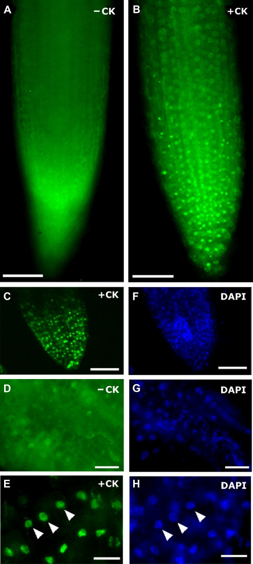
Figure 2.
Bacterial RR PhoB translocates to plant nuclei in root cells in response to HK activation with exogenous cytokinin. (A, B) Cellular localization of PhoB-GFP in roots of transgenic Arabidopsis plants. (A) Before cytokinin treatment, PhoB-GFP fluorescence appears diffused and throughout the cells. (B) After exogenous cytokinin treatment, the same root shows PhoB-GFP accumulation in sub-cellular compartments. (C–H) Detail views of roots (D, G) before and (C, E, F, H) after treatment with cytokinin showing that before cytokinin is applied, GFP fluorescence is diffused; after cytokinin exposure, the compartments in which PhoB-GFP accumulates (C, E) also stain with DAPI (F, H), indicating that they are nuclei (arrowheads). −CK, tissue before cytokinin treatment; +CK, tissue after cytokinin treatment; DAPI, tissues treated with DAPI to stain DNA. Scale bars, 50 μm in (A–C, F); scale bars, 10 μm in (D–E, G–H).

