Figure 5.
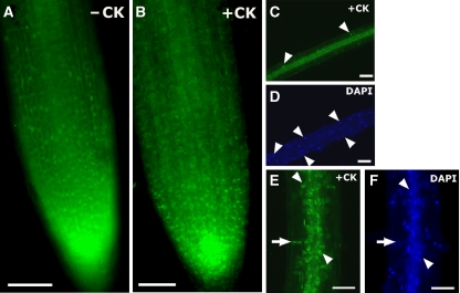
Cellular localization of mutagenized PhoBD53A-GFP in roots of transgenic Arabidopsis plants. (A) Fluorescence from PhoBD53A-GFP is diffused in an untreated root. (B) The same root showing PhoBD53A-GFP localization after cytokinin treatment. (C, D) Detailed view of a root showing that nuclear localization of PhoBD53A-GFP is variable and sporadic (arrowheads point to nuclei). (E, F) Detail view of another root showing that PhoBD53A-GFP accumulates at the base of cortical cells (arrows). Some nuclear localization of PhoBD53A-GFP can be seen in the root vascular tissue (arrowheads). −CK, tissues before cytokinin treatment; +CK same tissue after cytokinin treatment; DAPI, tissues treated with DAPI to stain DNA. Scale bars, 50 μm in (A, B); scale bars, 10 μm in (C–F).

