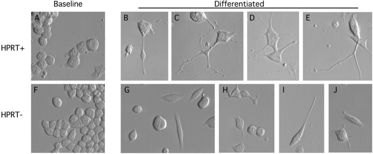
Figure 3.
Morphological appearance of parent and HPRT− mouse MN9D sublines. The parent MN9D cells are shown in the top row, and the HPRT− mutants are shown in the bottom row before (A and F) and after (B–E, G–J) sodium butyrate differentiation. Undifferentiated parent MN9D (A) and mutant (F) cultures appeared predominantly as simple round or ovoid cells. Differentiated parent cells demonstrated cell cycle arrest, enlargement and flattening of the soma, and elaboration of multiple complex branching neurites that often extended out of the field of view (B–E). Differentiated mutant cells demonstrated cell cycle arrest with soma changes similar to control cells. However, there was a high proportion of cells with no neurites (G and H), and the neurites tended to be short and simple (H–J).

