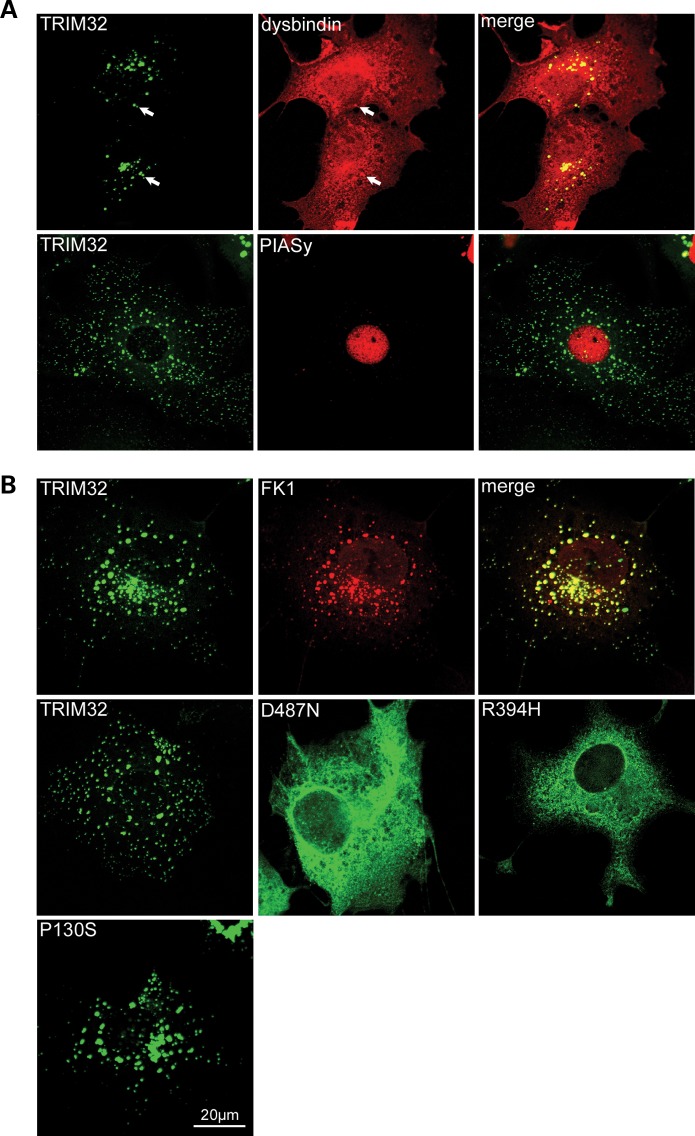
Figure 3.
Subcellular distribution of TRIM32, dysbindin and PIASy in COS-7 cells. (A) Co-localization of TRIM32–EYFP and dysbindin in COS-7 cells. TRIM32–EYFP and dysbindin were transfected into COS-7 cells and stained for dysbindin using PA3111A (red). Dysbindin is diffusely expressed in the cytoplasm and nucleus. In addition, dysbindin can be found in aggregates that co-localize with the TRIM32–EYFP fluorescence (arrows), which appear yellow in the merged image. Myc-tagged PIASy and TRIM32–EYFP were transfected into COS-7 cells and stained for myc using 9E10 (red). PIASy exhibited a nuclear localization, with TRIM32 residing in cytoplasmic speckles. (B) Localization of TRIM32–EYFP and mutants in COS-7 cells. COS-7 cells were transfected with TRIM32–EYFP, D487N–EYFP, R394H–EYFP and P130S–EYFP. TRIM32 and P130S form discrete cytoplasmic speckles, whereas D487N and R394H are predominantly cytoplasmic. TRIM32–EYFP transfected COS-7 cells were stained with FK1, which detects polyubiquitinated proteins (red). Merging the two images shows that the two antigens predominantly co-localize indicating that TRIM32–EYFP cytoplasmic speckles are associated with polyubiquitinated proteins.

