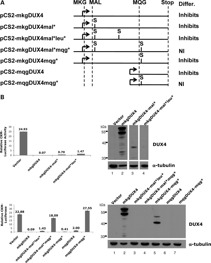
Figure 5.
Inhibition of myogenesis in the absence of DUX4 protein. (A) Schematic of the coding regions and stop-codon placements of the expression constructs tested. The methionines in an open reading frame with DUX4 are depicted with the two following amino acids (MKG, MAL, MQG) and the DUX4 translation stop codon indicated by STOP. S shows the regions where we have introduced a new translation stop codon. The column labeled Differ. indicates whether the expression of the indicated vector inhibited C2C12 differentiation (Inhibits) or did not inhibit differentiation (NI). (B) C2C12 cells transiently transfected with pCkm-luc and CMV-beta-galactosidase together with the indicated DUX4 expression. Bar graphs show luciferase activity relative to beta-galactosidase activity 24 h after induction in differentiation medium and western shows abundance of protein containing the epitope recognized by the 9A12 monoclonal antibody to DUX4. The two major bands in the western represent translation initiation at the MKG and the MAL methionines and they migrate at their predicted size. Note that the monoclonal antibody does not recognize in vitro translated mqgDUX4 (data not shown) indicating that the epitope is not contained within this region of the protein.

