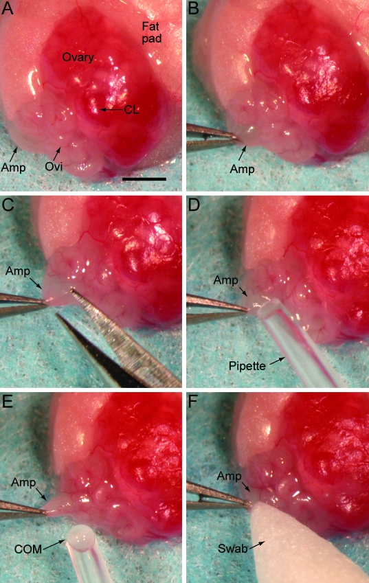
Figure 2.
Photographs of the surgical oocyte retrieval (SOR) process. (A) The ovarian fat pad, ovary, and oviduct are exteriorized and positioned as shown. (B) The ampulla is grasped by using Dumont forceps and held in place. (C) A 0.1- to 0.2-mm incision is made in the ampulla wall by using Vannas microdissecting scissors. (D) The cumulus oocyte mass (COM) is suctioned gently from the ampulla by using a gel-loading pipette attached to a mouth pipette. (E) The COM is not aspirated into the bore of the pipette tip but adheres to the tip once freed from the ampulla. (F) A swab is used to apply tissue adhesive to the ampulla incision. Amp, ampulla; CL, corpus Luteum; Ovi, oviduct; COM, cumulus oocyte mass. Bar, 1.0 mm.

