
Figure 9.
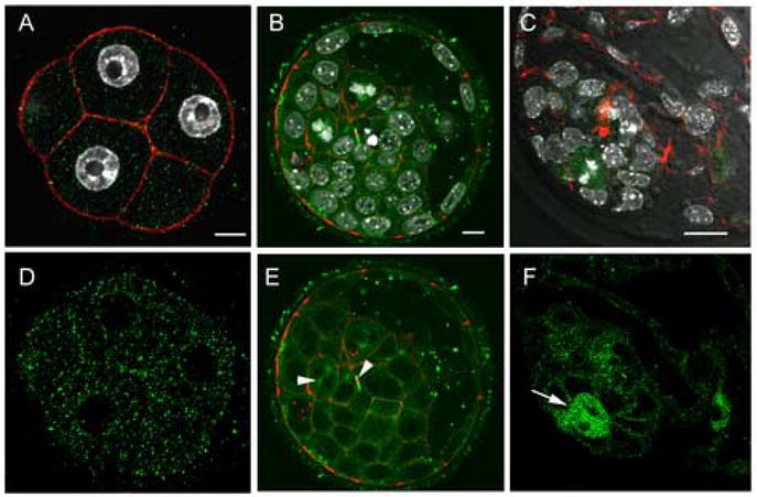
Distribution of activated SFKs in the morula and blastocyst. Embryos were fixed at the compaction (~48h post-hCG) and the expanded blastocyst (120h post-hCG) stages, then labeled with the clone-28 antibody (green) as well as Alexa-568-Phalloidin (red) to visualize f-actin. At the 8-cell compacted stage, activated SFKs were no longer associated with the nuclear envelope and were evenly distributed throughout the cytoplasm. In blastocyst stage embryos (Panel B,C,E,F), activated Src-family PTKs were concentrated at the cortex of most inner cell mass and trophoblast cells as well as at mitotic spindles and midbodies (arrow heads). At higher magnification, (Panels C, F) activated Src-family PTKs were concentrated in cells actively undergoing mitosis (arrows). Magnification is indicated by the white bar which represents 10μm.