
Figure 3. NK cells are developmentally normal in the absence of selectins.
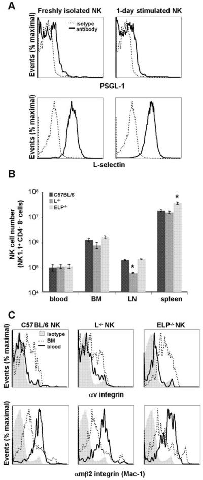
A. NK cells were isolated from spleens of C57BL/6 wt mice and either assayed directly or cultured overnight in activating medium containing IL-2 and IL-15. C57BL/6 NK cells were analyzed by FACS for expression of L-selectin and selectin ligand PSGL-1. B. NK cell numbers in blood (∼100μL), bone marrow (1 femur), lymph node (1 inguinal) and spleen of selectin-replete and selectin-deficient mice were determined from total cell number in the abovementioned tissues multiplied by percentage of NK1.1+, CD3- cells as determined by FACS. Data were obtained from at least 3 mice of each genotype and are presented as average cell numbers ± SEM; statistical significance for selectin-deficient mice as compared to selectin-replete mice within each organ has been calculated; *, p<0.05 by two-tailed Student’s t test. C. Freshly isolated blood and bone marrow collected from C57BL/6 selectin-replete and selectin-deficient mice were analyzed by FACS for expression of αV integrin and CD11b (Mac-1; αMβ2 integrin). As expected, αV integrin expression decreases on blood NK cells as compared with the immature NK cells in the bone marrow, while CD11b expression increases.