
Figure 4. NK cells from selectin-deficient mice are functional in vitro.
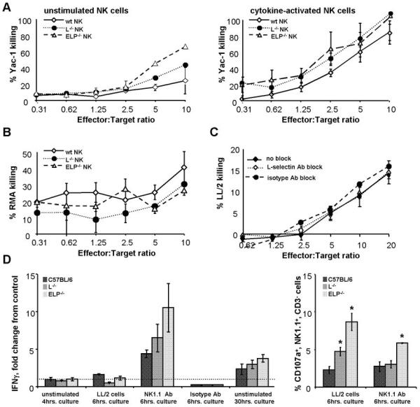
A, B. NK cells were isolated from spleens of selectin-deficient and control C57BL/6 mice. Purified NK cells were co-cultured with varying ratios of tumor target cells in a standard cytotoxicity assay. Cytotoxicity of either freshly isolated NK cells (A, left panel) or NK cells activated overnight in medium containing IL-2 and IL-15 (A, right panel; B, C) was determined. Target killing was measured using the Promega CytoTox 96® Non-Radioactive Cytotoxicity Assay. NK cells from selectin-deficient mice (A, B), as well as NK cells treated with L-selectin-blocking antibody MEL14 or an isotype control antibody prior to and during the assay (C), displayed normal ability to kill tumor targets. D, left panel: IFNγ secretion by purified control and selectin-deficient NK cells was assayed by ELISA. NK cells were either left in culture medium with no additional stimulation for 4hrs. after isolation, stimulated by co-culture with LL/2 tumor cells for 6hrs, stimulated by NK1.1 antibody or an isotype control antibody for 6hrs, or allowed to remain in culture medium for 30hrs. Data are normalized to IFNγ secretion by control NK cells cultured with medium alone for 4hrs. Selectin-deficient NK cells do not demonstrate any statistically significant differences from selectin-replete C57BL/6 NK cells within each group. D, right panel: in addition, NK cells stimulated by LL/2 tumor cells or by the NK1.1 antibody for 6hrs. were assayed for degranulation by CD107a FACS analysis. All analyses were performed for at least 3 animals of each genotype. Results are expressed as bar and line graphs, error bars = SEM
*, p<0.05 by two-tailed Student’s t test.