Figure 3.
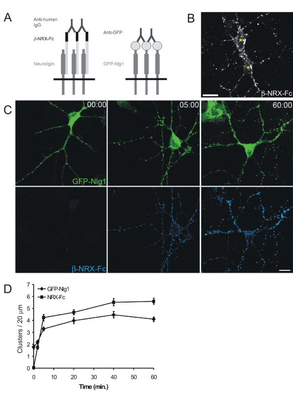
Patching of endogenous and recombinant Nlg. (A) Diagram depicting the two methods of patching: pre-oligomerized β-NRX-Fc fusion proteins were used to patch endogenous Nlg and anti-GFP antibodies to patch recombinant GFP-Nlg1. (B) Fluorescence image of live cortical neurons demonstrating β-NRX-Fc bound to non-transfected neurons (asterisks indicate cell bodies). Scale bar, 20 μm. (C) Confocal images of cortical neurons transfected with GFP-Nlg1 and immunostained with antibodies against GFP (green) and human Fc (blue) after 0, 5 and 60 minutes of patching with β-NRX-Fc. Scale bar, 20 μm. (D) Quantification of the time course of patching for both β-NRX-Fc clusters and GFP-Nlg1 clusters per 20 μm of dendrite (n ≥ 13 neurons for each time point).

