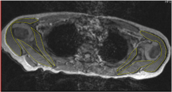
Figure 1.
FISP acquisition MRI in axial plane showing affected and normal contralateral shoulder. In the affected left [L] shoulder there is a biconcave glenoid form (type 3) and humeral head subluxation. The contralateral [R] shoulder is normal. Measured areas of infraspinatus, subscapularis and deltoid are outlined.

