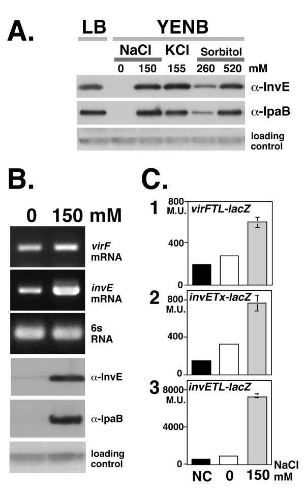
Figure 1.
A. InvE and IpaB expression in different osmotic conditions. An overnight culture of strain MS390 at 30°C was inoculated into fresh YENB medium with or without osmolytes and then incubated at 37°C until mid-log phase (A600 = 0.8). Medium, osmolyte, and concentration are indicated at the top of the panel. Antibodies used for detection are indicated on the right of the panels. A cross-reactive unknown protein detected by the anti-InvE antiserum was used as a loading control for InvE Western blot analysis throughout this study. B. Expression of >invE and virF mRNA and InvE and IpaB protein expression in S. Sonnei. Total RNA (100 ng) and 10 μl of the indicate culture were subjected to analysis of mRNA and protein levels, respectively. The 6S RNA ssrS gene was used as control for RT-PCR. Primers and antibodies are indicated on the right side of the panels. Concentration of NaCl in the medium is indicated at top of the panel. C. Expression of invE and virF >promoter-driven reporter genes. Wild-type S. sonnei strain MS390 carrying the indicated reporter plasmids were subjected to a β-galactosidase assay: Graph 1, virFTL-lacZ translational fusion plasmid pHW848; Graph 2, invETx-lacZ transcriptional fusion plasmid pJM4320; Graph 3, invETL-lacZ translational fusion plasmid pJM4321. Concentration of NaCl is indicated at the bottom of the graphs. Details of the control experiments, indicated by black bars (NC)are described in methods.

