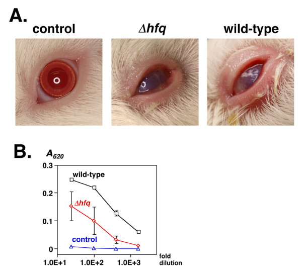
Figure 6.

A. Development of experimental keratoconjunctivitis. Photograph of the left eyes of guinea pigs 4 days after infection. A bacterial cell suspension (5 × 10⁸ cells) was dropped into the conjunctival sacs of male Hartley guinea pigs, and the animals were observed for four consecutive days. Left panel, control animal infected with LB medium alone; middle panel, animal infected with Δhfq strain MS4831; right panel, animal infected with wild-type strain MS390. B. Serum antibodies against effector molecules of TTSS. Sera were obtained from three animals two weeks after infection. Serial 25-, 100-, 400-, and 1600-fold dilutions were added to immobilized soluble effector molecules (see Methods) on a microtiter plate. Antibodies were detected using peroxidase-conjugated anti-guinea pig IgG. The absorbance at 620 nm (A620) of each well was monitored after the addition of ABTS using a microplate reader. Black squares, animals infected with wild-type strain MS390; red diamonds, animals infected with Δhfq strain MS4831; blue circles, control animals that received LB medium. Data represents the means and standard deviation of 2 samples.
