
Figure 5. Effect of TGF-β1 treatment on caspase-3, ERK1/2, AKT and SMAD2 activation in colorectal cancer cell lines deprived from anchorage.
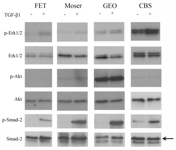
Immunoblot assay results for caspase-3, ERK1/2, AKT and SMAD2 and their active forms in FET, Moser, GEO, and CBS maintained in suspension for 20 hours with exogenous TGF-β1 (10ng/ml) are shown. Activation of SMAD2 is present in all of the cell lines, but only FET and CBS display increased ERK1/2 phosphorylation after TGF-_1 treatment.