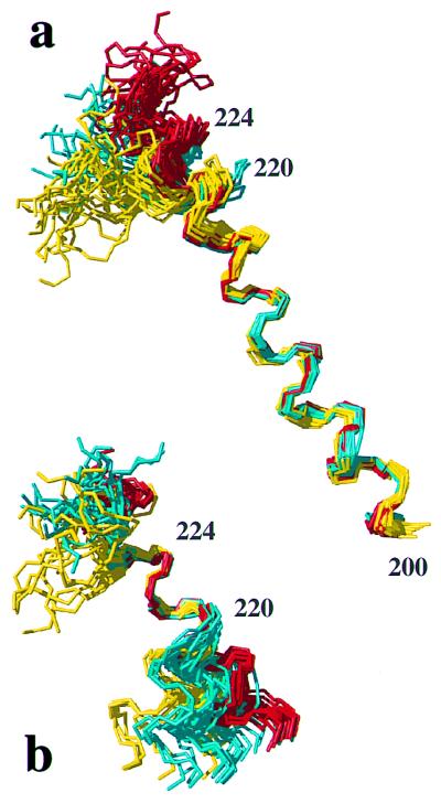
Figure 4.
Comparison of helix 3 in hPrP(121–230) (red), mPrP(121–231) (yellow), and hPrP(R220K) (cyan) with the NMR structures represented by bundles of 20 conformers (28). (a) Polypeptide backbone of residues 200–229 superimposed for best fit of the residues 200–218. (b) Residues 213–229 superimposed for best fit of the residues 221–225.

