Fig. 1.
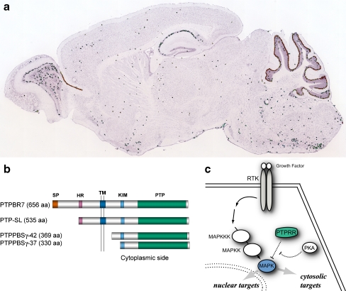
PTPRR isoforms are expressed in the brain and interact with MAPKs. a PTPRR mRNA levels in adult mouse brain. A sagittal section demonstrating high transcript levels in the hippocampal area and most notably cerebellar Purkinje cells, extracted from the Allen Brain Atlas [72]. b Schematic representation of the different mouse PTPRR protein isoforms. Protein names and lengths, in amino acid residue numbers (aa), are on the left. SP signal peptide, HR hydrophobic region, TM transmembrane region, KIM kinase interacting motif, PTP protein tyrosine phosphatase domain. c MAPK localization in the cytoplasm and into the nucleus depends on the balance between growth factor activation and KIM-containing PTP inhibition. Activated PKA will prevent the PTPRR–MAPK association by phosphorylation of the KIM domain [34]. RTK receptor tyrosine kinase, MAPKKK MAPKK kinase, MAPKK MAPK kinase, MAPK mitogen-activated protein kinase

