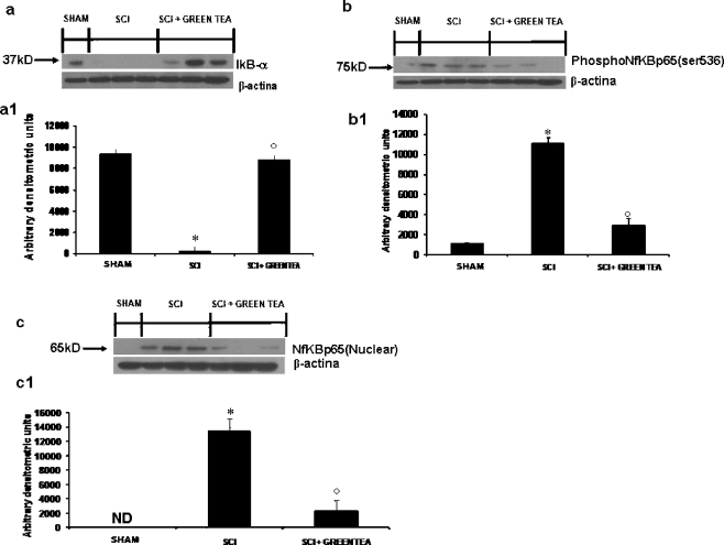
Fig. 3.
Effects of GTE treatment on IkB-α degradation, phosphorylation of NF-kB subunit p65 on Ser 536 and nuclear NF-kB p65. By Western blot analysis, a basal level of I kB-α was detected in the spinal cord from sham-operated animals (a, a1), whereas in SCI control mice, I kB-α levels were substantially reduced (a, a1). GTE treatment prevented the SCI-induced I kB-α degradation (a, a1). In addition, SCI caused a significant increase in the phosphorylation of Ser536 at 24 h (b, b1). The treatment with GTE significantly reduced the phosphorylation of p65 on Ser536 (b, b1). Moreover, the levels of the NF-kB p65 subunit protein in the nuclear fractions of the spinal cord tissue were also significant increased at 24 h after SCI compared with the sham-operated mice (c, c1). The levels of NF-kB p65 protein were significantly reduced in the nuclear fractions of the spinal cord tissues from animals that had received GTE treatment as shown in c (c1). Immunoblotting in a–c are representative of one spinal cord of five analysed. The results in a1, b1 and c1 are expressed as mean ± SE mean from five blots. *p < 0.01 versus Sham, degree symbol p < 0.01 versus SCI

