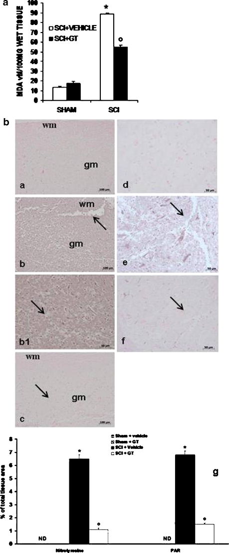
Fig. 6.
Effect of GTE on lipidic peroxidation, nitrotyrosine formation and PARP formation. The MDA levels were significantly increased in the spinal cord tissues from SCI operated mice (a). GTE reduced the spinal cord injury-induced increase in MDA levels (a). Spinal cord section from sham-operated mice did not stain for nitrotyrosine (b, a). In addition, section obtained from SCI animals demonstrated positive staining for nitrotyrosine mainly localised in inflammatory cells and in the nuclei of Schawnn cells in the wite and gray matter (b, b, b1). GTE treatment reduced the degree for positive staining for nitrotyrosine in the spinal cord tissue (b, c). Moreover, immunoistrochemistry for PAR revealed the occurrence of positive staining for PAR in mice subject to SCI (b, e). GTE treatment reduced the degree for positive staining for PAR in the spinal cord tissue (b, f). No positive staining for PAR was observed in the tissue section from sham-operated (b, d). Densitometry analysis of immunocytochemistry photographs (n = 5 photos from each sample collected from all mice in each experimental group) for nitrotyrosine and PAR (b, g) from spinal cord tissues was assessed. The assay was carried out by using Optilab Graftek software on a Macintosh personal computer (CPU G3-266). Data are expressed as percent of total tissue area. Values shown are mean ± SE mean of ten rats for each group. wm white matter, gm gray matter. This figure is representative of at least three experiments performed on different experimental days. *p < 0.01 versus sham, e, degree symbol p < 0.01 versus SCI.

