
Figure 3.
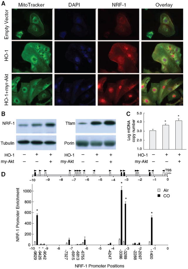
HO-1 increases nuclear NRF-1 and Nrf2 occupancy of NRF-1 ARE promoter motifs. Active HO-1 increases NRF-1 expression and nuclear protein by confocal microscopy in HL-1 cells (A). Cotransfection with HO-1 and active Akt containing an amino-terminal myristoylation signal (myr-Akt) produces a further 3-fold increase in nuclear NRF-1 protein (A and B) accompanied by 8- to 10-fold increases in Tfam expression (B, right) as well as mtDNA copy number (C). A search of the NRF-1 promoter for Nrf2-binding sites 10 kb upstream of the TSS identified multiple ARE motifs investigated by ChIP (D). Arrows indicate orientation of each ARE motif relative to the ARE consensus. Gray boxes are motifs meeting the consensus sequence and black boxes show active motifs. The graph shows DNA enrichment by Nrf2 ChIP at each NRF-1 ARE motif. Values along the abscissa indicate positions of DNA regions amplified by qPCR. Black bars depict significant DNA enrichment after DCM/CO at positions −1400, −3289, 3386, and −9829. Nrf2 binding increased 11 to 13-fold at each site. Values represent the means±SEM of independent experiments performed in triplicate (*P<0.05).