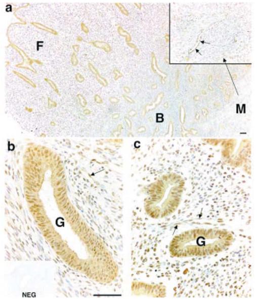
Figure 2.
Immunohistochemical localisation of PGIS in human endometrial tissue (functional layer and basal-myometrial junction) collected in the proliferative phase of the menstrual cycle. Strong staining was detected in the glandular epithelial cells (G) in basal (B) and functional (F) layers (a-c) and in smooth muscle cells in the myometrium (M) (enlarged area in inset to a). Stromal staining was detected in the basal (b) and the functional layer (c) and endothelial cell reactivity was present in blood vessels (indicated by arrows) in all layers. NEG, negative control; scale bars = 50 μm.

