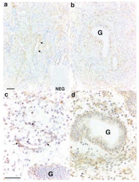
Figure 3.
In situ hybridisation of IP receptor in sections of human endometrial tissue collected in the proliferative phase of the menstrual cycle. IP receptor was expressed in smooth muscle cells in the myometrium (a) and reactivity was also present in glandular epithelial cells (G) and in stromal cells in both the basal layer (b) and the functional layer (c and d). IP receptor was also expressed in vascular endothelial cells (indicated by arrows in a and c). NEG, negative control; scale bars = 50 μm.

